Although hydronephrosis is one of the most commonly detected prenatal anomalies, it poses challenges to urologists because of the variability of the condition and because the medical community has not achieved consensus about when testing and treatment are needed, or about which tests and treatments are appropriate.
At Children’s Health℠, Children’s Medical Center Dallas, our FETAL Center (Fetal Evaluation and Treatment Alliance Center) and urologists have worked together to evaluate and/or treat hundreds of cases of hydronephrosis. And we regularly consult with outside physicians to help them assess the condition’s severity and decide on appropriate monitoring and treatment. These consultations have taken on an added dimension during the COVID-19 pandemic, via a new TeleFETAL program that enables us to virtually consult with physicians and families from afar.
“With hydronephrosis, it can be easy to over-test and overtreat because there’s no clear guidance or best practices,” says Craig Peters, M.D., Division Director of Pediatric Urology at Children’s Health and Professor of Urology at UT Southwestern. “We’re here to help physicians identify the cases that are worrisome and determine how to manage them, and to counsel families – and TeleFETAL is making that easier than ever.”
Detecting and assessing dilation prenatally
Hydronephrosis cases are typically referred to our FETAL Center after kidney dilation is detected in a standard prenatal ultrasound at 18 to 20 weeks. The FETAL Center then turns to Dr. Peters and his colleagues, who review the imaging, order additional tests if needed, and evaluate whether future intervention could be warranted.
“The FETAL team works very closely with the Department of Urology to facilitate a seamless transition from prenatal to postnatal care, and the child’s pediatrician, an obstetrician and a maternal fetal medicine specialist are fully integrated along the way,” Dr. Peters says.
Our urology team bases its evaluation on five factors:
Degree of dilation
Nature of the kidney tissue (parenchyma)
Location of the dilation
Parts that are dilated
Amount of amniotic fluid
“All this information guides how we assess the condition, how we counsel the parents based on concern for potential problems and the potential need for an intervention – and how often we want to see babies after they’re born,” Dr. Peters says.
Our urology team first looks at kidney dilation, using the Society for Fetal Urology’s grading system to score the dilation (zero to four). Mild cases – about half of all cases – show little dilation, and some can resolve without treatment. In more severe cases, calyces appear dilated. With grade four, all the calyces are dilated, and the kidney tissue appears stretched thin.
“When we see dilation, our evaluation and monitoring concentrate on whether it might affect kidney function over time,” Dr. Peters says.
Our team also uses imaging to look at the kidney tissue. With increased echogenicity, the kidneys will look brighter than the liver or spleen.
“That can mean the kidney is being stressed,” Dr. Peters says. “In more severe cases, you may see cysts – sometimes a lot with extreme obstruction – and that's a sign of poor kidney function and might mean the kidney isn’t developing.”
As part of the evaluation, it’s also important to note the point where the dilation stops.
“It's like a dam in a river,” says Dr. Peters. “If just the kidney is dilated, then the blockage is typically at the beginning of the ureter. If you can see dilation down to the bladder, the second most common site of blockage, that's ureterovesical junction obstruction.”
Bladder dilation can be hard to see in an ultrasound. If both kidneys and both ureters are dilated, the bladder may also be obstructed. In boys, bladder outlet obstruction can indicate posterior urethral valves, which can damage the kidneys and even be lethal.
If there is hydronephrosis with reduced amniotic fluid, Dr. Peters and his colleagues will look for a bladder outlet obstruction, which can put the pregnancy at risk.
How to monitor hydronephrosis in newborns
After birth, there is no general consensus about how monitoring and treatment should proceed. Most babies with hydronephrosis appear fine, and there is no single test to measure urinary tract obstruction. Even a kidney function test may not reveal severe obstruction in one kidney if the other is healthy.
As a solution, Dr. Peters recommends an ultrasound two to six weeks after birth.
“Right after birth, babies typically are very dehydrated and aren’t making a lot of urine,” he says. “So hydronephrosis that showed up prenatally may not show up again for a few weeks. That can create confusion. If a baby does receive imaging within the first 48 hours, we would recommend repeating the ultrasound a few weeks later and having us review it directly.”
Diagnostic tests for hydronephrosis
If babies continue showing dilation after the first postnatal ultrasound, Dr. Peters may recommend two diagnostic tests, depending on the character of the dilation.
A bladder X-ray or a voiding cystourethrogram (VCUG) can detect reflux and diagnose bladder obstruction. Dr. Peters advises a VCUG if the ureters are dilated, if both kidneys in a boy are dilated, if the bladder looks abnormal, or if there is a duplicated system.
However, Dr. Peters recognizes that the use of VCUG is controversial. Reflux can cause an infection, sometimes suddenly, which can cause significant illness in a neonate. Not all children with dilation will have reflux and some will do fine without making the diagnosis, since reflux will resolve on its own in many children. However, the test itself can cause infection in about 2-3% of patients.
“I usually will counsel parents, either prenatally or postnatally, about VCUG’s pros and cons, and about why we might not need it,” Dr. Peters says. “If we proceed with the test, I will put the baby on a low dose of antibiotic to prevent infection.”
If one or both kidneys are dilated to a significant degree, Dr. Peters and his colleagues use a second test, the MAG-3 – diuretic renogram – to evaluate the severity of obstruction and the effects on kidney function.
When to consider surgery for hydronephrosis
Either at baseline or follow-up, Dr. Peters recommends surgical repair if the hydronephrosis is worsening, if function for a kidney is reduced 10-15% from normal, or if the patient develops pain or gets a urinary infection that affects the kidney.
“At that point, hydronephrosis will not usually go away on its own,” Dr. Peters says. “And sometimes, when we’ve been monitoring a baby for a few years, the parents decide not to wait any longer and to just go ahead with surgery.”
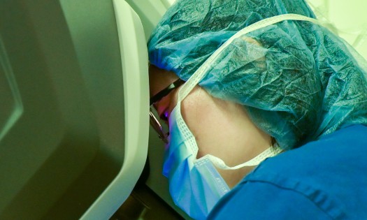
Our team uses pyeloplasty, usually by robotic-assisted laparoscopy, to repair the most common cause of ureteral obstruction, ureteropelvic junction obstruction. We’re one of the only urology departments in the world where every surgeon is certified in robotic surgery, and the procedure has a 98% success rate with a shorter recovery time.
Obstruction of the ureter at the level of the bladder (ureterovesical junction obstruction, or obstructive megaureter) are less common, and we typically address them via megaureter repair. This procedure is often done using open surgery, by removing the section of the ureter that is blocked and then reattaching the ureter to the bladder. This surgery can be done robotically as well. After surgery, the team monitors the children with ultrasound or the MAG-3 to ensure satisfactory drainage.
“The decisions are not simple, the children require ongoing monitoring, and there are risks to any approach, non-operative or operative,” says Dr. Peters. “But we always do our best to weigh those risks vs. the potential benefits.”
The benefits of telemedicine for hydronephrosis
Our TeleFETAL program has brought key advantages for families after a diagnosis of hydronephrosis, by enabling them to have a video call with Dr. Peters or another one of our urologists. We use this call to explain the baby’s condition, walk through future tests and potential treatments, and answer the family’s questions. This spares families a trip to our Dallas campus, while still giving them the chance to meet their baby’s providers before birth.
TeleFETAL also can enable us to advise community physicians and their patient families who may not need further treatment, or the stress and travel that can come with it. For example, we were recently referred a woman who was told that prenatal imaging showed a severe problem. She lived in Lubbock – more than 300 miles from Dallas – and expected her baby to be transferred to Children’s Health after delivery for dialysis.
“First, we met with the mother over TeleFETAL and reviewed the imaging, in collaboration with Dr. Matthias Wolf from the Division of Pediatric Nephrology,” says Dr. Peters. “We advised them that the condition was not as severe as initially thought and that her baby could receive whatever care it needed close to home. I connected her with a colleague in Lubbock, who saw the baby on day one. The tests were reassuring, and the baby’s doing well. It was much easier for mom – and a remarkably beneficial use of telemedicine with a good outcome.”
Learn more about innovative urology care and research at Children’s Health


