Prenatal detection of congenital heart disease (CHD) is an essential step in providing the highest quality of care for patients born with CHD. Prenatal detection of CHD can be challenging for many reasons. Small cardiovascular structures, a rapid heartbeat and normal movements inside the womb can make it hard to get clear images. Fetal echocardiography also has limiting factors that hinder accurate results, such as the mother’s build, fetal position, low amniotic fluid and fetal bone development.
The Children’s HealthSM Fetal Heart Program has long been a leader in overcoming these challenges to accurately detect CHD prenatally. The team performs hundreds of echocardiograms annually and is one of the leading centers in North Texas. Now, Children’s Health is one of only a few centers in the United States to offer fetal cardiac magnetic resonance imaging (fetal CMR) as a complement to fetal echocardiography.
Fetal CMR is an advanced diagnostic imaging test that can supplement fetal echocardiography in the prenatal diagnosis of congenital heart disease. The limitations of fetal echocardiography become apparent in complex vascular cardiac anatomy, late gestation diagnoses and with challenging maternal acoustic windows. Fetal CMR can overcome these obstacles, in addition to providing information about distributions of fetal blood flow through various cardiac pathologies.
“We’ve been studying fetal CMR as part of our research protocols to better understand its advantages compared to other imaging tests and how to get the best results using it,” says Catherine Ikemba, M.D., Pediatric Cardiologist and Director of the Fetal Heart Program at Children’s Health and Professor at UT Southwestern.
Advantages of fetal CMR
The advantages of fetal CMR are that it doesn’t require sedation for either the mother or baby, and that it’s best performed during the third trimester of pregnancy when there is much less fetal movement.
“This is the timeframe when it can be harder to successfully see things on a fetal echocardiogram, so diagnosis or prognostication can be challenging. With fetal CMR, the bigger the fetus, the better,” says Mansi Gaitonde, M.D., Pediatric Cardiologist at Children’s Health and Assistant Professor at UT Southwestern.
Fetal CMR is performed with a unique MR safe/compatible Doppler ultrasound device to gate the fetal heartbeat and appropriately time image acquisition based on the cardiac cycle of the fetal heart. The device goes on the mom’s abdomen and connects directly to the MR scanner.
“We’re one of a few centers that have this device, and we’ve refined the way we use it to get very clear, high-quality images,” says Munes Fares, M.D., Pediatric Cardiologist at Children’s Health and Assistant Professor at UT Southwestern.
By better assessing blood flow patterns and visualizing arterial anatomy, fetal CMR also has the potential to better characterize the presence of certain diagnoses that are very challenging to make by fetal echocardiography, such as coarctation of the aorta or pulmonary atresia with aortopulmonary collaterals.
Bringing together experts to perform and interpret fetal CMR
The fetal CMR team at Children’s Health consists of pediatric cardiologists, pediatric radiologists, cardiac MRI technologists, nurses and a physicist who work collaboratively to get accurate results while delivering the best experience for pregnant mothers.
“We’re one of only a handful of pediatric heart centers to have a dedicated physicist who develops and refines these innovative sequences and can assist real-time while scanning the mother,” says Dr. Fares. Given the small size of the fetal heart, unique MR and physics concepts must be applied to accurately visualize the fetal cardiac and extracardiac anatomy and function.
“In addition to building a team with a depth of expertise in this technology, we’re also dedicated to being very attentive to moms who may be anxious about having the test,” says Dr. Fares.
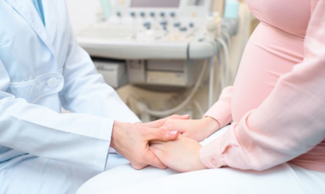
Before the scan begins, a nurse ensures that the mother is in an optimal position. The MRI techs assess placental and fetal positioning and guide the test while ensuring maternal comfort. Pediatric cardiologists are present the whole time and provide direct feedback to the fetal heart team.
Whole-patient care for families facing CHD
After CHD is diagnosed, parents also receive supportive services from a broader multidisciplinary team at the Children’s Health Heart Center and UT Southwestern including cardiology, maternal fetal medicine, neonatology, cardiothoracic surgery, intensive care, social work, psychology and the palliative care team.
“This broader team helps plan and prepare for the baby’s life after birth. We also actively address the most common maternal complication of pregnancy, perinatal mental health disorders. A prenatal diagnosis of congenital heart disease can be devastating for a family. We screen for anxiety, depression, and post-traumatic stress on our Dallas campus. Our heart center psychologists and social workers provide resources for psychological support and mental health care,” says Dr. Ikemba.
Promising outcomes in fetal CMR research
The Children’s Health team is collaborating with the Society for Cardiovascular Magnetic Resonance and the Fetal Heart Society to better understand the capabilities of this new diagnostic tool. One exciting possibility is that fetal CMR can help better predict prognosis in some types of CHD.
“Fetal CMR enables us to better understand the distribution of blood flow in a fetus and may visualize cardiac and extracardiac structures better when imaging by fetal echo is limited. With this new understanding, we hope to establish better algorithms for what is normal and abnormal in fetal life with time. Ultimately, the hope is that we can become better at predicting certain types of severe CHD that have proven challenging by fetal echocardiography time and time again,” Dr. Gaitonde says.
These severe types of CHD include:
Hypoplastic left heart syndrome with restrictive septum
Tetralogy of Fallot or pulmonary atresia with multiple aortopulmonary collateral arteries
“We’re also exploring how fetal CMR can evaluate the relationship between the heart, brain and placenta with regards to blood flow and oxygenation. This could help us better understand the connection between CHD and other organ issues beyond the immediate postnatal period – and help with family counseling and postnatal planning. Fetal CMR is now available for clinical ordering by our fetal heart and maternal fetal medicine teams when patient specific questions in fetal CHD arise,” Dr. Gaitonde says.
Dr. Ikemba adds, “If we can tell the difference between those who are going to be critically ill in the first hour of life, versus those who just need attention in the first days or weeks of life, we've made a huge impact. Improving the lives of these families is what drives us to pursue cutting-edge research and technology that will raise the bar on detection rates and ultimately lead to life-changing outcomes.”
Learn more about advanced fetal imaging at Children’s Health >>


